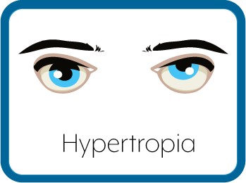
Hypertropia (Vertical Strabismus)
What is Hypertropia?

In general, strabismus (or tropia) is defined by frequency (intermittent or constant), laterality (right, left, or alternating), and direction (horizontal or vertical).
Horizontal strabismus is termed esotropia (inward turn of the eye) or exotropia (outward turn of the eye). Vertical strabismus is termed hypotropia (downward turn of the eye) or hypertropia (upward turn of the eye).
Therefore, Hypertropia is a form of vertical strabismus where one eye is deviated upwards in comparison to the other eye.
Causes of Hypertropia
The most common cause of hypertropia is palsy (weakness) in one of the cranial nerves, the third or fourth nerve. Hypertropia may also co-exist with infantile strabismus, esotropia or exotropia. Other causes of hypertropia include problems that may be congenital (present at birth) or develop later:
- 3rd or 4th cranial nerve palsy
- Brown syndrome: Problem with a tight eye muscle tendon
- Duane syndrome: Problem with wrong innervation of the eye muscles
Signs and Symptoms of Hypertropia
Hypertropia may be intermittent (happening occasionally) or constant, and the symptoms may be barely noticeable. The most common symptoms are:
- One or both eyes wandering upward
- Head tilt to compensate for the eye misalignment
Those with Hypertropia may also be experiencing the following, whether they are aware of it or not.
Suppression
Suppression is not perceived by the patient, but rather by the observer. Suppression happens when the visual system suppresses the image coming from the deviated eye. The amount of suppression, which can vary from small suppression scotomas in binocular fusion to large suppression areas on the affected side and amblyopia, depends on various factors such as the size of the strabismus and age of onset.
Diplopia
Diplopia is the medical term for double vision. In the case of a hypertropia, the diplopia is vertical.
Confusion
Vision confusion occurs when two images are perceived in the same location, due to a misalignment of retinal correspondence points on the fovea. This symptom is rare, when compared to diplopia.
Tests Used to Assess Hypertropia
Hirschberg Test
A light source located in front of the optometrist's eyes is directed at the patient's eyes, while the patient is asked to fixate the light source directly. The corneal light reflex is observed. The Hirschberg test is considered normal when the corneal light reflexes are slightly decentred nasally (about 5º, due to angle kappa). In the case of a hypertropia, the light reflex of the deviated eye is located below the light reflex of the fixing eye. The amount of deviation can be grossly estimated by multiplying the mm of deviation by 15PD.42,43
Krimsky Test
This test uses prisms to complement the Hirschberg test. Prisms are positioned in front of the deviating eye, base-down in the case of a hypertropia, and are progressively increased until a neutral Hirschberg test is obtained. It is particularly helpful in patients that don’t collaborate well in the former test, especially with low visions.
Cover/Uncover Test
The cover/uncover test allows for the diagnosis of tropias, when performed correctly. In order to achieve this, the optometrist needs to briefly cover the eye that is fixing and see if there is a refixation movement of the fellow eye. In the case of a hypertropia, the non-fixing eye moves downward as it takes up fixation. If no refixation is observed the other eye may be the fixing one, in which case it is covered and the test is performed again. It is very important for the cover to be very brief, since a prolonged cover will break binocular fusion and provoke a possible phoria that can be misinterpreted as a -tropia. A pure -phoria won’t have a positive cover/uncover test, while a -tropia is also associated with positive alternate cover test.
Simultaneous Prism Cover Test
A test that can be used to estimate the angle of deviation attributable to a -tropia. The amount of PD that have to be added in order to revoke refixation movements in the deviating eye, correspond to the deviation angle. This shouldn’t be confused with the alternating prism cover test for the correction of a -phoria component, in which case binocular fusion is interrupted. In the case of incomitant strabismus due to muscle paresis or restrictive syndromes, one prism is placed over the eye with limited ductions to measure the primary deviation and a second prism is placed in front of the good eye to measure secondary deviation. The deviation is always bigger when the eye with limited ductions is fixing (i.e. the prism is over the normal eye)
Worth 4 Dot Test
Allows for the diagnosis of diplopia and suppression. A different light filter is positioned in front of each eye, a green and a red light filter. The patient is asked to look at 4 different dots: 2 green dots on either side; one red dot on top; one white dot on the bottom, forming a cross. Green dots can only be seen by the eye with the green light filter and the red dot can only be appreciated by the eye with the red filter, while the white dot can be seen by both eyes. If the patient sees 5 lights instead of 4, a diplopia is present. If the lights seen by one eye are below the expected position, it means that eye is hypertropic (the image is projected on the superior retinal quadrants, which perceive the lower visual fields). If the patient sees less than 4 lights, suppression is present.
Red Filter Test
Same principle as the Worth dot test, but with only one light source and one light filter (red) in front of the eye to be examined. The patient seeing a pink light is a normal test result. If two lights are perceived, diplopia is present. If the patient only sees a white light, suppression of the eye with the red filter is present.
Maddox Rod Test
One Maddox rod is positioned in front of each eye, while the patient is asked to look at a light source. This way, each eye will only see one linear streak of light. In order to test for a vertical tropia, the Maddox rods have to be placed in order to create streaks at 180º degrees. If one streak is perceived below the other, a hypertropia/phoria is present. Since the Maddox rod test is highly dissociative, it doesn’t allow for a differential diagnosis between a -phoria and –tropia.
Bagolini Striated Lens Test
This test is very similar to the Maddox rod test, with the exception that Bagolini striated lenses allow for a better view of the peripheral visual field, giving more binocular clues. This way, less dissociation is present and a better distinction between a small suppression scotoma with peripheral fusion and a large suppression scotoma is possible.
Treatment of Hypertropia
Treatment for hypertropia aims to ensure proper vision in both eyes and aligning the eyes. Common treatment options include:
- Glasses to correct vision problems such as nearsightedness, farsightedness, or astigmatism that may be contributing to the hypertropia. This may include the use of prism lenses.
- Vision Therapy, again often in conjunction with glasses or prism lenses.
- Eye patch over the strong eye to improve the vision in the weak eye.
- Surgery on the eye muscles to realign the eyes.
The Vision Wiki
- Lazy Eye
- History of Lazy Eye
- Convergence Disorders
- Convergence Insufficiency
- Exophoria
- Esophoria
- Medical Terms
- Accommodation Disorders
- Accommodative Esotropia
- Accommodative Insufficiency
- Amblyopia
- Lazy Eye in Adults
- Strabismic Amblyopia
- Refractive Amblyopia
- Anisometropia
- Critical Period
- Strabismus
- Anomalous Retinal Correspondence
- Transient Strabismus
- Exotropia
- Esotropia
- Hypotropia
- Hypertropia
- Eye Problems
- Binocular Vision
- Physiology of Vision
- Lazy Eye Treatments
- Reading
- Fields of Study
- Research
- Glaucoma
- Virtual Reality
- Organizations
